- • Explore how DNA and chromosomes replicate and divide during mitosis and meiosis
- • Understand the differences between mitosis and meiosis
- • Reinforce what happens during each step of mitosis and meiosis
Instructions: Keep this open in your browser window and type what you need, you can print when you're ready. You can find more labs and get your biology A game at learn.biobuddy.com.
Learning Objectives
Motivation
Dr. Nystrom
Lead Scientist
The goal of this lab is to give credit to how COOL mitosis and meiosis are.
As a graduate student at The University of Virginia, I had the excellent opportunity to teach biology, anatomy, and physiology to all grade levels of UVA students. These students were very high performing, and many planned to pursue graduate degrees themselves, with goals ranging from PhDs to medical school to dental school to public health.
Given their high level of academic performance, there were certain subjects that I assumed would be easy for them. One such subject was when the class covered cell cycle - mitosis and meiosis. Cell cycle is something that most students have been learning since middle, or even elementary school! I figured we wouldn't have to spend much time on it.
What I learned when we arrived at the cell cycle unit is that most students had a very strong base level understanding of mitosis and meiosis, but often their understanding lacked depth. I came to realize that even my own understanding of mitosis and meiosis lacked depth!
This led me to think that since we learn about mitosis and meiosis from such a young age, we assume, as I did, that it is simple and does not require much time. We learn the basics and move on. But I think this is a disservice to our learning and understanding of the cell cycle.
THUS, for this unit activity we will actually be doing a visualization activity to reinforce, step by step, what happens during mitosis and meiosis, understand how they are different, and hopefully develop a better understanding of why the cell cycle is SO interesting and important.
Background
A deeper dive into meiosis and mitosis can be found on Biobuddy - Cell Cycle (AP Bio - Unit 4.5), Regulation of Cell Cycle (AP Bio - Unit 4.6), and Meiosis & Genetic Diversity (AP Bio Unit 5.1 + AP Bio Unit 5.2)
NOTE - For the purpose of this activity, we are specifically talking about eukaryotic cells and cell cycle.
The cell cycle is the process by which cells maintain and reproduce themselves, and proper progression of the cell cycle is crucial for the functioning and growth of a healthy organism! Moreover, in the case of meiosis, proper progression of the cell cycle is critical for the maintenance and well-being of entire populations and species!
The cell cycle is made up of 2 main stages - Interphase and M phase, or mitosis, and each of these stages has phases during which specific and important processes take place!
We will get into the nitty gritty of interphase, mitosis, and meiosis in the following activity!
Lab Activity
For this activity, we will go through mitosis and meiosis step by step, taking notes on what happens at each stage. This is an activity that my pre-med, undergraduate students at UVA struggled with, so I'm hoping you all won't find yourselves in that same position!
The Steps of the Cell Cycle
As we discussed before, there are two main steps of the cell cycle! Interphase and M phase. Label the phases of Interphase below:
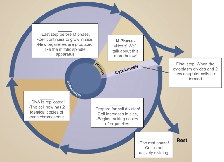
Watch video 4.5 - Cell Cycle for a more in-depth explaination of this illustration
Illustrate what happens to the cell as it progresses through the cell cycle!
Below is your starting cell. For the purpose of this activity, we will only illustrate what happens to the chromosomes and general cell size.
*Draw - The nucleus with 2 sets of homologous chromosomes. These are unreplicated chromosomes, so they should look like 2 sets of lines of the same length, but different colors. The same length represents them being the same chromosome, the different color represents them not being identical*
Starting Cell (Before Interphase)
Interphase
To me, interphase feels like preparing for a big event, like a big party! Interphase is the phase during which a cell prepares for cell division. Just like people preparing for an event, cells spend most of their time in interphase, with only a small amount of time spent in the M phase. There are three stages of interphase - G1 (Gap 1), S (Synthesis), and G2 (Gap 2).
G1 - What happens to the cell during G1?
*Draw - The cell as it appears above. Then, draw the cell after G1. It should be a slightly larger version of the same cell.*
Starting Cell
Cell after G1
S - What happens to the cell during S phase?
*Draw - The cell as it appears after G1, then the cell after S phase. The cell after S phase looks like the same cell, but with the DNA replicated. So an identical copy of each of the 4 chromosomes.*
Cell after G1
Cell after S phase
G2 - What happens to the cell during G2?
*Draw - The cell as it appears after G2 Phase. The cell should be a bit bigger, the nucleus should contain the replicated chromosomes.*
Cell after S phase
Cell after G2 Phase
Mitosis
To me, mitosis feels like the big event, and I think many of us perceive it that way. While the preparation (interphase) is actually completely necessary, we spend more time talking about and learning about mitosis. And it is kind of a big event! Mitosis is when the cell actually divides up the organelles and DNA, and a new cell is made! Below you will illustrate what happens to the DNA as the cell progresses through mitosis.
Prophase
Cell during Prophase
Prophase
- Replicated chromosomes becomes visible
- Sister chromatids (identical copies of a chromosome) form the X chromosomal structure
- Prometaphase
- The nuclear envelope begins to disintegrate
Draw the above in the cell to the left
Metaphase
Cell during Metaphase
Metaphase
- Condensed chromosomes line up along the center of the cell.
- Helpful hint - During Metaphase, chromosomes Meet at the middle.
- Spindle fibers come out from the centrioles and attach to the centromere of the chromosomes.
Draw the above in the cell to the left
Anaphase
Cell during Anaphase
Anaphase
- Sister chromatids are pulled apart by spindle fibers
Draw the above in the cell to the left
Telophase
Cell during Telophase
Telophase
- Nuclear envelopes reform with chromosomes inside of them.
- Chromosomes begin to decondense.
- The new cells are not quite separated yet!
Draw the above in the cell to the left
- Each cell should have 4 chromosomes and look identical to the starting cell.
- The resulting cells are diploid.
Cytokinesis
Cell after Cytokinesis
Cytokinesis
- Cytoplasm divides
- Cell membrane forms
Draw the above in the cells to the left
- Each cell should have 4 chromosomes and look identical to the starting cell.
- The resulting cells are diploid
Meiosis - Meiosis I and Meiosis II
Meiosis is a specialized version of the cell cycle that produces gametes. It is during meiosis that cells divide to create new eggs and sperm. While mitosis produces 2 identical daughter cells, certain steps of meiosis introduce genetic variation into the DNA, therefore producing daughter cells with distinctly different genetic material. This variation in genetic material is imperative for the survival of a species and is the basis of genetic adaptation and evolution. There are 2 main parts of meiosis, meiosis I and meiosis II, and each have their own steps.
Meiosis I
Prophase I
Cell during Prophase I
Prophase I
- Replicated chromosomes becomes visible
- Sister chromatids (identical copies of a chromosome) form the familiar chromosomes structure that looks like an 'X'
- Homologous chromosomes pair up in tetrads
- Crossing over occurs during Prophase I
Draw the above in the cell to the left - Make sure to include a visual representation of crossing over
Crossing Over:
- Crossing over Is an incredibly important part of meiosis!
- This is when homologous chromosomes exchange genetic material
- THIS IS WHERE GENETIC VARIATION IS INTRODUCED!
Metaphase I
Cell during Metaphase I
Metaphase I
- Condensed chromosomes line up along the center of the cell
- Spindle fibers come out from the centrioles and attach to the centromere of the chromosomes
Draw the above in the cell to the left
Anaphase I
Cell during Anaphase I
Anaphase I
- Homologous chromosomes are pulled apart by spindle fibers
- Sister chromatids stay together
Draw the above in the cell to the left
Telophase I
Cell during Telophase I
Telophase I
- Homologous chromosomes move to opposite sides of the cell
- New nuclear envelopes may form in new sides of the dividing cell
- Cytokinesis begins to divide the cytoplasm of the cell into 2 new cells
- The new cells are not quite separated yet!
Draw the above in the cell to the left
- Each side should have 1 set of sister chromatids for each of the homologous chromosomes!
Meiosis II
Meiosis II is very similar to meiosis I, with 2 important exceptions.
- The DNA is not duplicated before meiosis II
- There is no crossing over in meiosis II
- The final product of meiosis II is 4 haploid cells
Telophase I
Cell during Telophase I
Telophase I
- See above!
Draw - the same thing you drew for telophase I above
Prophase II
Cell during Prophase II
Prophase II
- Cytokinesis ocurrs
- DNA is not replicated between meiosis I and meiosis II
- Each new cell should have one set of replicated sister chromatids from each homologous pair, for a total of 2 sets.
Draw the above in the cells to the left
Metaphase II
Cell during Metaphase II
Metaphase II
- The sets of sister chromatids line up along the metaphase plate
- Spindle fibers come out from the centrioles and attach to the centrosomes of the sister chromatids
Draw the above in the cells to the left
Anaphase II
Cell during Anaphase II
Anaphase II
- The spindle fibers pull the sister chromatids apart, pulling 1 chromosome from each pair opposite sides of the cell
Draw the above in the cells to the left
Telophase II
Cell during Telophase II
Telophase II
- Nuclear envelope begins to reform around chromosomes
- Spindle fibers disappear
- Cytokinesis begins
- The new cells are not quite separated yet!
Final Product
Final Product - 4 Haploid Cells
Discussion
- Cell cycle and cell division are incredibly popular subjects of study in modern research. Why do you think researchers care so much about cell division?
- What could happen if there are flaws in the cell cycle?
- Can you think of any diseases that are associated with changes in cell cycle or cell division?
- Why is crossing over so important?
- In the introduction of this lab, I said - "proper progression of the cell cycle is critical for the maintenance and well-being of entire populations and species." Why do you think I am making this claim?